Anatomage Table 13 Update: The Full List of Added Content and Features (2026)
Built on more than 15 years of continual refinement, this latest iteration of the Anatomage Table software provides a deeper way to explore the complexity and multidimensionality of the human body. With every release, Anatomage continues to introduce increasingly detailed anatomical content, functional physiology, and clinical applications designed to support medical education and clinical training.
Anatomage Table 13: New Content and Features
Release date: 2026
New Content and Tools
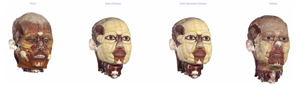
Adrian
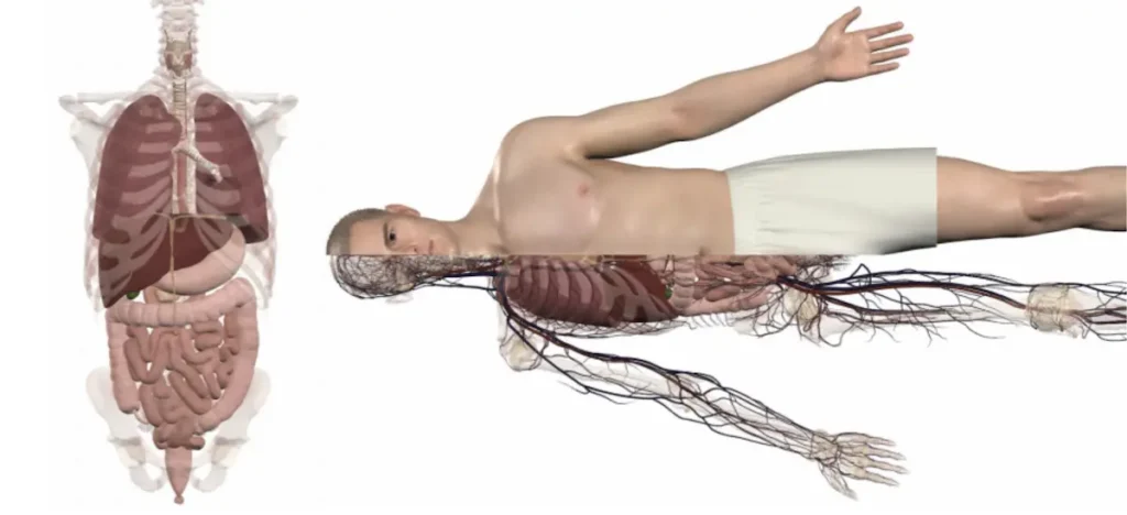
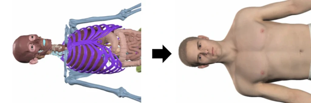
Adrian, the male counterpart to Ariel, was modeled from Anatomage’s real 3D cadaver data to present a healthy and standardized textbook-friendly model in anatomical position. Adrian offers students a unique opportunity to begin their anatomical studies with a 3D body that closely mirrors textbook illustrations.
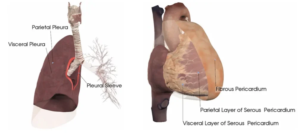
The Adian and Ariel models contain highly specific, accurate anatomy. Visualize intricate structures such as pleura, meninges, fascia, pericardial layers, fat pads, and detailed reproductive anatomy.
Obese and Severely Obese Ariel
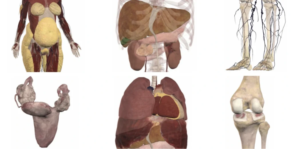
These two new models were created to enhance medical education regarding the anatomical changes produced by obesity. These reference bodies represent Class I and Class III obesity, following WHO and CHC classifications. See the “Cadaver Information” folder on the Table desktop for more details.
Obese: Class I Obesity (BMI 30-34.9)
Skin changes (folds, stretch marks, and cellulitis)
Increased subcutaneous and visceral fat volume, with resulting organ displacement, compression, and reduced pulmonary volume
Increased severity of all changes in the Obese model
Enlarged ovaries with large number of immature follicles
Atheriosclerosis (aorta) and cardiomegaly
Pancreatic and kidney hypertrophy
Liver Hypertrophy and Steatohepatitis (MASH)
Varicose veins
Severe osteoarthritis (knees)
Brain atrophy
New Features and Tools
Reset Button
Within the Orientation menu, there is now a full reset feature. This icon will reset all features of a scan back to the initial conditions, including positioning, structure visibility, colors, labels, and dissections.
Anatomy Construction Activity
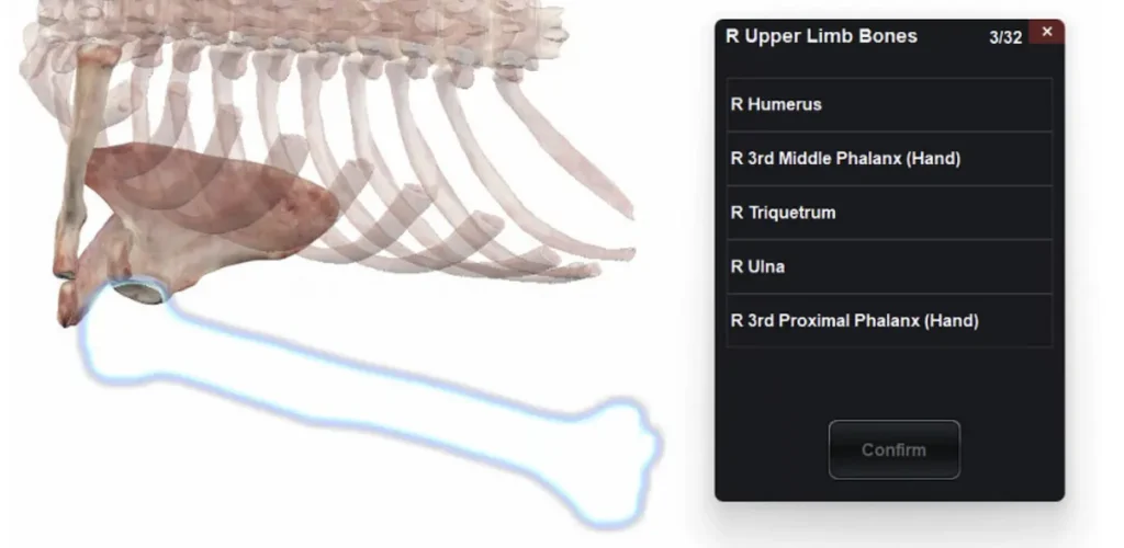
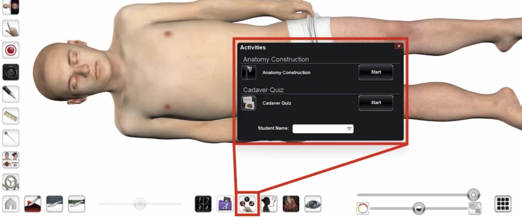
The Quiz Mode feature has expanded to include a new style of activity: Anatomy Construction. In an Anatomy Construction activity, students are challenged to build anatomical pathways in the correct sequence, reinforcing spatial relationships. Includes pre-built quizzes.
DICOM Image Quiz Activity
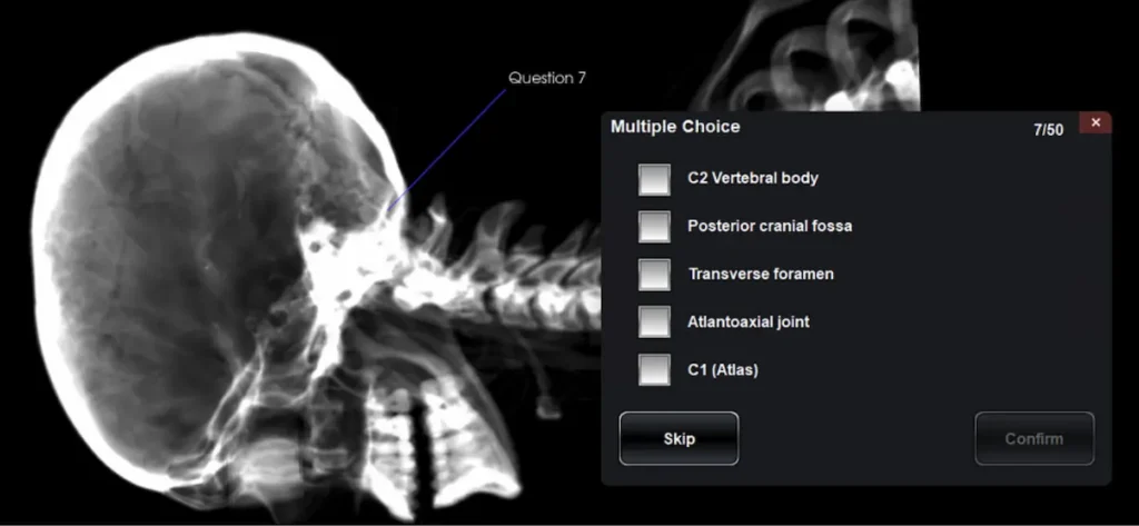
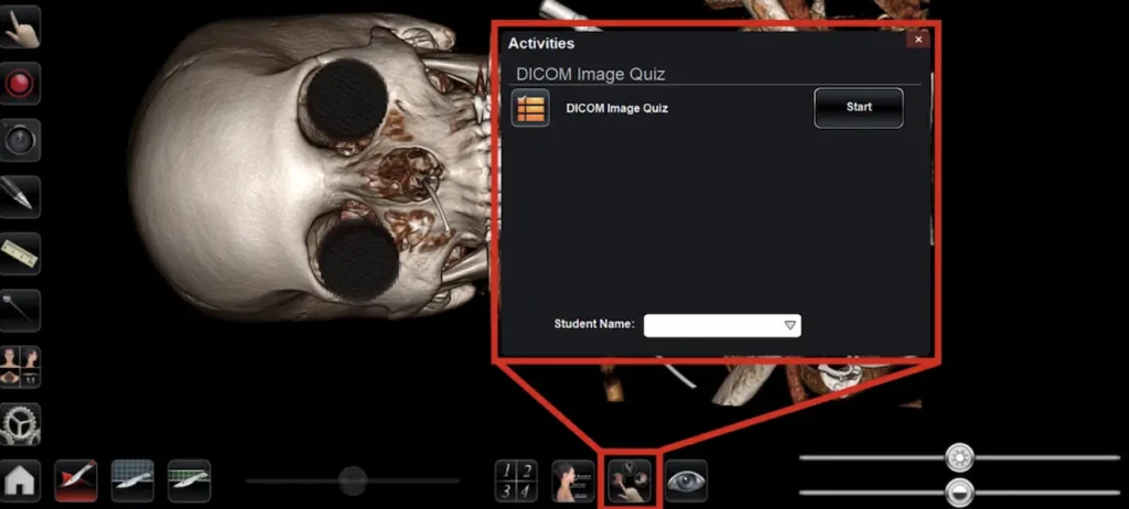
This feature utilizes 3D reconstructions of CT scans to challenge students to identify structures and sections in a real patient scan. DICOM Image Quizzes test anatomical identification in a clinical imaging context, and allow for customization of views and filters.
Locate the DICOM Image Quizzes through the new “Activities” icon in the main menu, or by selecting the “Activities” icon from within a Case Library scan.
Slab Clipping Tool
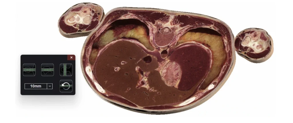
The new slab tool automatically creates parallel clipping planes to isolate a cross-sectional slab of the body.
Case Library: HiP-CT Scans
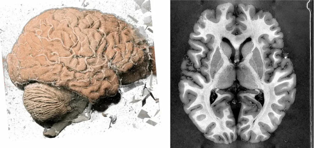
17 Hierarchical Phase-Contrast Tomography scans have been added to the Table. Originating from the Human Organ Atlas repository, these scans have been enhanced with Anatomage’s 3D rendering technology to deliver medical imaging at an extraordinary level of detail.
Feature Improvements
Updated Quiz Mode (Activities)
New Name: The “Quiz Mode” interface and icon have been updated and renamed. In Cadavers, Cadaver Quizzes and Anatomy Construction Activities can both be found under the “Activities” icon.
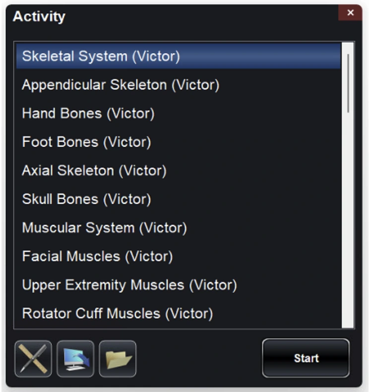
Premade Activities (Quizzes): Tap the “Start” button next to either Anatomy Construction or Cadaver Quiz to see a list of premade activities. Create your own Activities using the create icon and import existing quizzes into the Activity menu using either the VPF Import or Folder icons.
Schematics Flow
The existing cardiovascular schematics have been updated to include flow. This feature allows students to connect complex pathways to a simple diagram while simultaneously understanding the direction of blood flow.
Additional Bodies in Multi-Body
Additional bodies have been added to the Multi-Body tool. Comparisons can now be made between the virtual cadavers (Victor, Vicky, Hans, Hanna, High Res Female Head, and High Res Female Pelvis), as well as the Reference Bodies (Ariel, Adrian, Obese, Severely Obese). More than 200 ready-to-use scenes are pre-built into the software for easy comparison.
Changes to the Dissection Tool Icons
The advanced Dissection Tool icons have been updated for clarity.
Point-to-Point Dissection: Place two points and dissect through a custom cylinder between them.
Sculpt: Define a custom area and remove all structures from within that area.
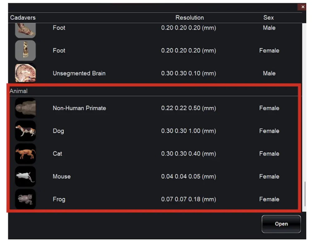
Animal Models Relocated
The interactive 3D animal models are now located at the bottom of the “Cadavers” menu underneath the “Animal” header. Animal CT and MRI scans can still be found in the Case Library underneath the “Animal” filter.
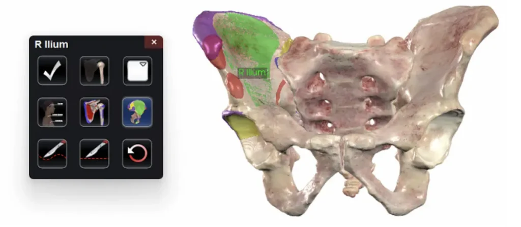
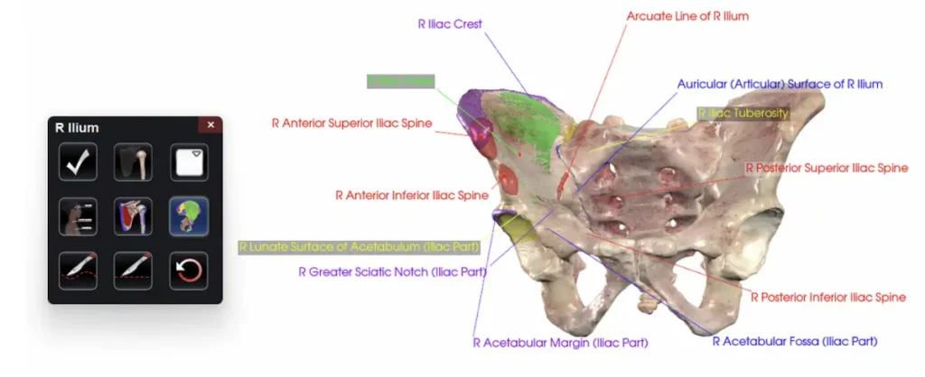
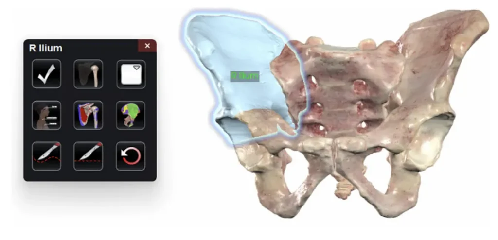
New Bony Landmark, Origin, & Insertion Behavior
When using the Bony Landmarks or Muscle Origin & Insertion tools, there is now a “three tap” behavior.
Tap 1: Turn on all the highlighted regions for the selected bone. No labels will appear. Tapping any highlighted region will turn on the annotation of that region.
Tap 2:Turn on all annotations for all highlighted regions for the select bone. Tapping any highlighted and annotated region will turn off the annotation of that region.
Tap 3: Turn off all the highlighted regions for the selected bone.
New Functional Anatomy
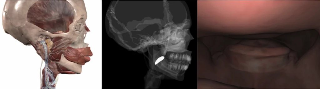
Swallowing
Visualize the complex muscular motions and neural stimulation involved in the highly-coordinated process of swallowing. With a 3D model on the left side, switch the right side between X-Ray and fluoroscopic views to emphasize the relevant anatomy while delivering a clinical perspective.
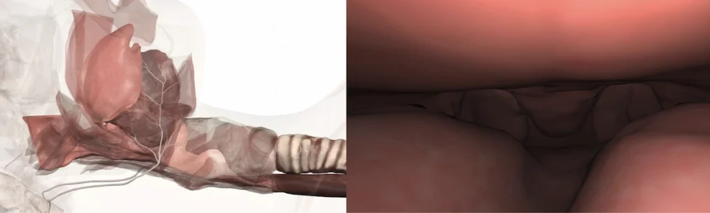
Sleep Apnea
Visualize the structures, movements, and neural stimulation involved during obstructive sleep apnea episodes. The simulation demonstrates airway collapse and reopening while connecting the episode to the role of the hypoglossal nerve.
In both Swallowing and Sleep Apnea, interact by playing and pausing the animation, and switch between fluoroscopic and internal views.
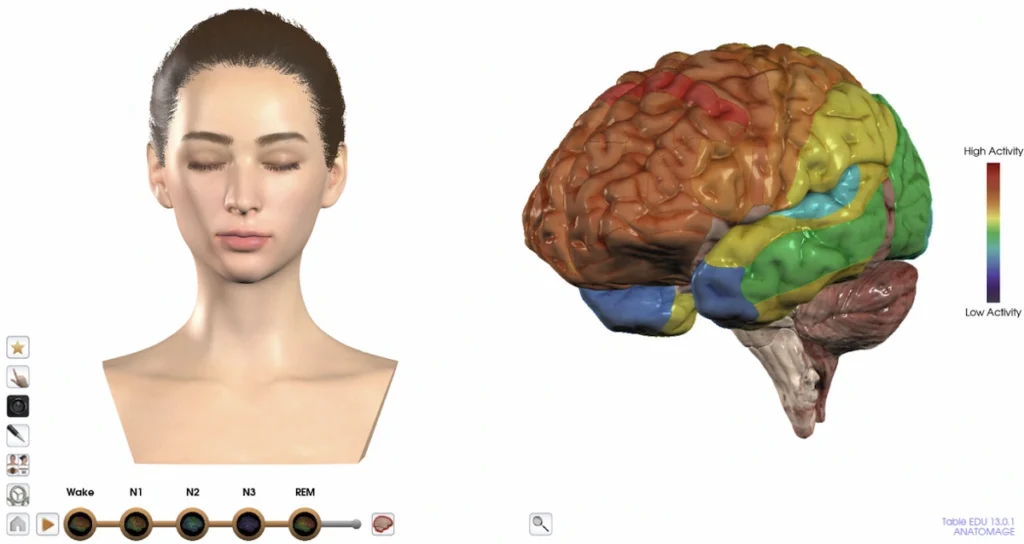
Sleep Physiology
Illustrate the changes in brain activity throughout the sleep cycle. Visualize how brain activity changes between N1, N2, N3, and REM sleep.
Explore the stages of sleep.
Identify the functions of Brodmann Areas in the brain. Visualize the difference in sleep under the influence of caffeine, insomnia, and sedatives.
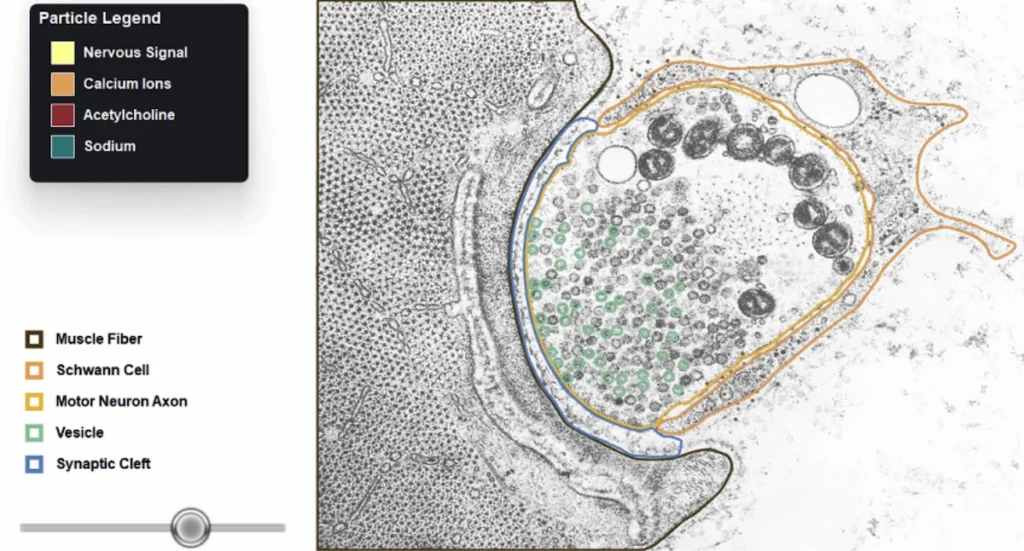
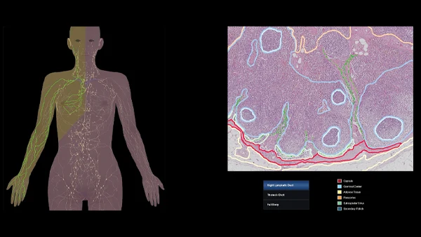
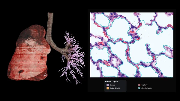
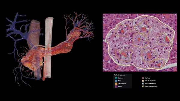
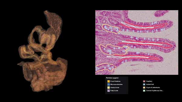
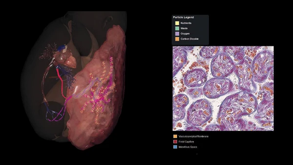
New 3D x Histology Simulations
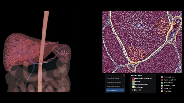
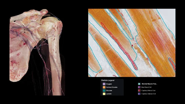
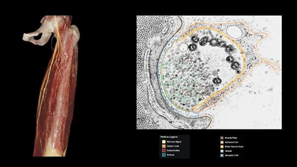
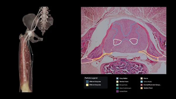
These simulations, similar to the existing Renal Physiology simulation, pair Anatomage’s real 3D anatomy with an animated histology slide to bridge the gap between anatomical structures and their microscopic physiological activity.
Play and pause particle flow animations in 3D and 2D histology environments.
Visualize key stages in physiological processes.
Toggle on detailed annotations of histology slides.
Lymphatic Physiology: Lymphatic drainage through vessels, nodes, and collecting ducts.
Alveolar Gas Exchange: O2 and CO2 diffusion across the alveolar membrane.
Hepatic Physiology: Protein, lipid, and carbohydrate processing, storage, and release. Bile, glucose, cholesterol, and lipoprotein synthesis. Bilirubin formation, transport, conjugation, and excretion.
Muscle Capillary Exchange: Delivery of O2 to skeletal muscle through arterial circulation and removal of CO2 and lactate through venous circulation.
Neuromuscular Junction: Action potential and synaptic transmission, including Ca2+ triggered acetylcholine release and Na+ influx.
Reflex Arc: Monosynaptic and polysynaptic reflex arcs with illustrated neural pathways.
Pancreatic Secretion: Glucose delivery to pancreatic β cells and Ca2+ dependent insulin release. Includes animation for hypoglycemia.
Intestinal Villi Absorption: Nutrient absorption along the small intestine, including monosaccharides, amino acids, and fatty acids.
Placental Exchange: Exchange of O2 , nutrients, CO2,, and metabolic waste between the maternal and fetal circulations.
We use cookies to ensure that we give you the best experience on our website. By continuing to use our website, you agree with our use of cookies in accordance with our privacy policy. You can reject cookies by changing your browser settings.OkPrivacy policy