Non-Human Primate Cadaver
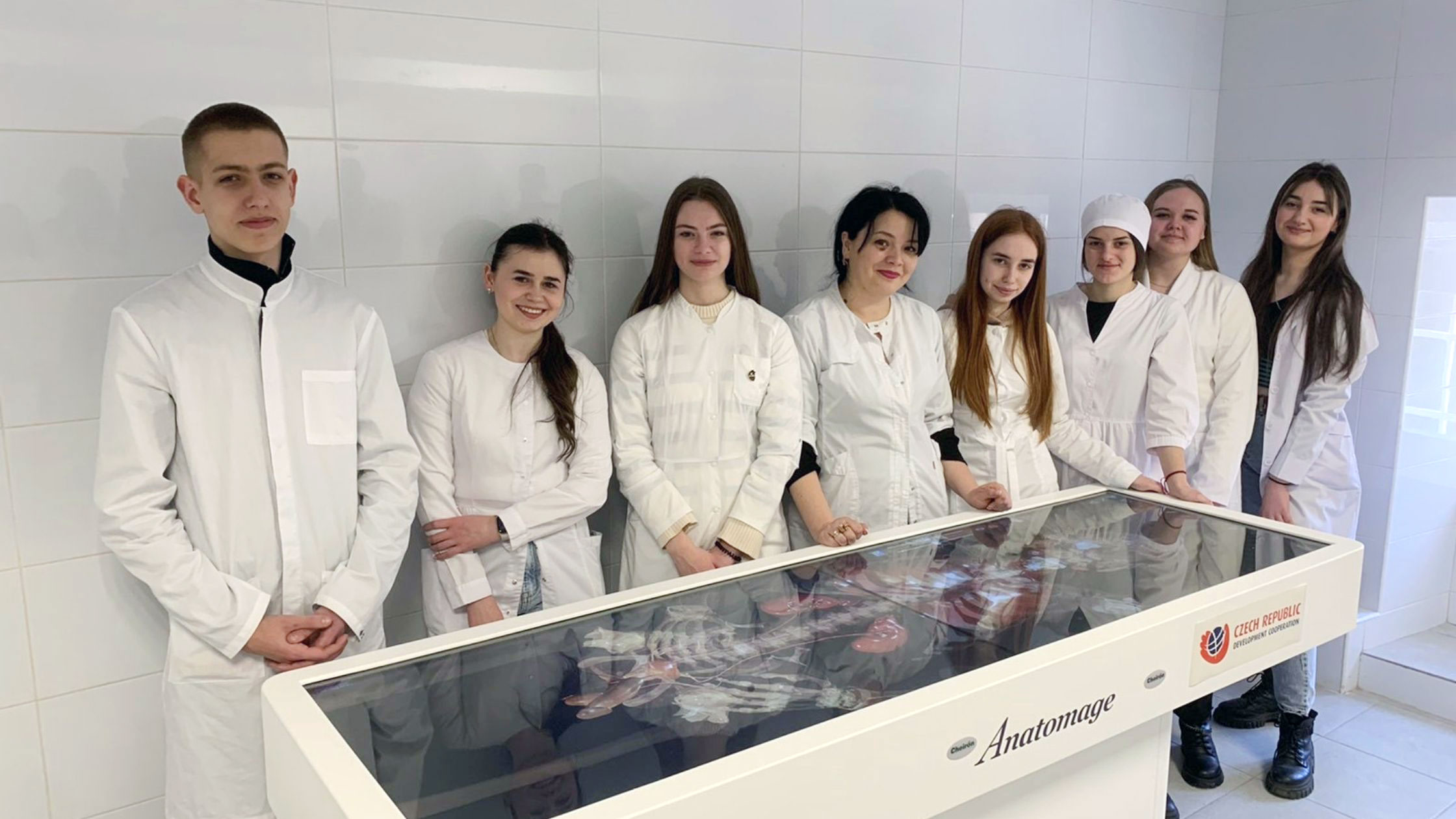
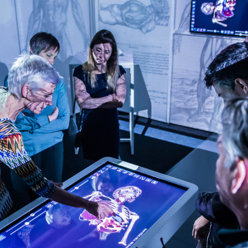
Available on the Anatomage Table, a state-of-the-art 7-foot touchscreen medical device, this digital model enables interactive and comprehensive exploration of non-human primate anatomy.
Traditional methods of studying primate anatomy are limited and can pose health risks. Anatomage’s digitized monkey offers a medically accurate, risk-free, and ethical solution, providing an authentic view of non-human primate anatomy like never before.
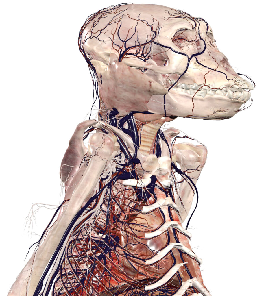
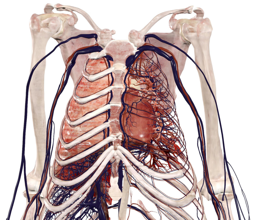
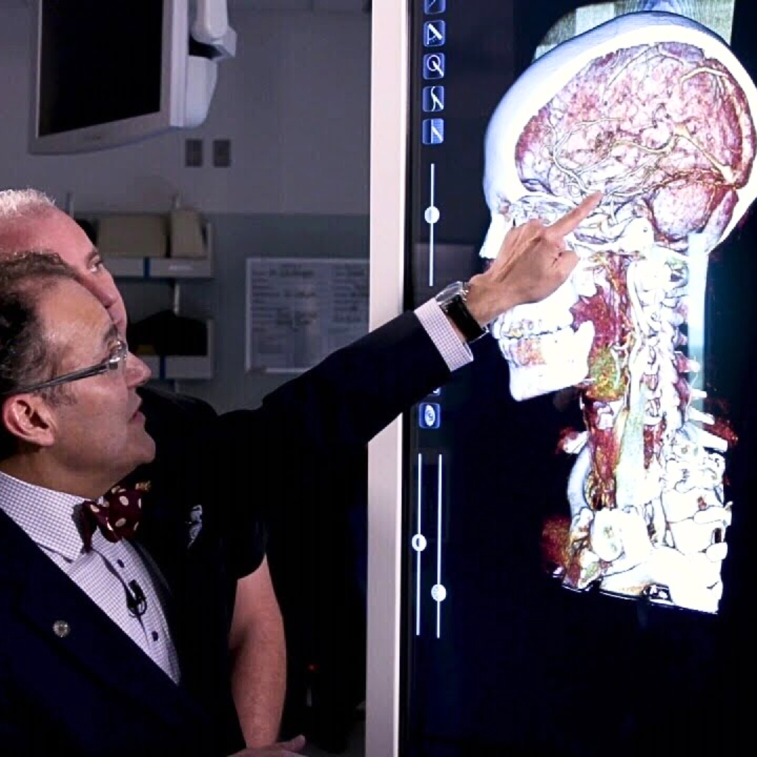
Anatomage has meticulously digitized the entire body of a female rhesus monkey, capturing thousands of segmented structures with precise annotations. The cardiovascular system, including veins and arteries, is fully connected and anatomically accurate. With a resolution down to 220 microns, the model provides a crisp, detailed visualization of intricate structures.
This digital primate cadaver is fully interactive on the Anatomage Table’s touchscreen interface, allowing students to explore the anatomy in 3D. Users can tap to identify specific structures, zoom in for close inspection of small bones and veins, and navigate through the primate’s complex anatomical landscape.
Anatomage’s technology embodies the 3Rs—Replacement, Reduction, and Refinement—by offering a groundbreaking, non-invasive approach to veterinary education. Real animal scans, rendered in exceptional detail, let students explore anatomy interactively, reducing the need for animal specimens while enhancing hands-on learning.
The digitized monkey model fully replaces traditional animal cadavers. It offers a realistic, detailed study experience without physical specimens. Students can explore complex primate anatomy safely, with no zoonotic risks. This model removes ethical concerns linked to using live or preserved animals. It recreates the value of cadavers in a sustainable, virtual environment.
Beyond the monkey model, the Anatomage Table features a library of digitized animal cadavers, including frog, cat, dog, and mouse. These virtual models provide a comprehensive, ethical alternative, significantly decreasing the need for live animals in educational dissection and training.
On the Anatomage Table, these digitized cadavers can be used to simulate a wide range of surgical procedures. The Table provides students with virtual scalpels and tools to perform various surgical and experimental techniques, such as craniotomy, ultrasound, and more. This approach eliminates the pain and distress associated with performing these procedures on live animals.
Anatomage Bodies provide significant benefits to medical education, supporting everything from anatomy visualization to clinical applications.
Ethical Dissection.
The Anatomage Table provides an ethical way to explore real animal anatomy without involving live animals, allowing students to build both scientific knowledge and compassion as they conduct biomedical research.
Sustainable Approach
Our digital dissection leaves no environmental footprint, offering students a safe way to explore anatomy without impacting the environment.
Detailed, Accurate, Interactive
Our digitized animal cadavers are modeled from real specimens, offering the highest level of accuracy and detail in anatomical visualization.
Read about how the Anatomage Bodies on the Anatomage Table provide significant benefits to the medical school community.
If you’re not an Anatomage Table user and would like to schedule a demo, please click the button below to get in touch with us.