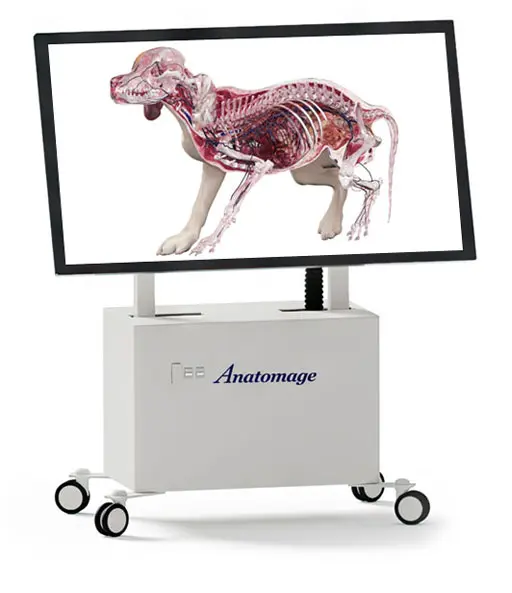
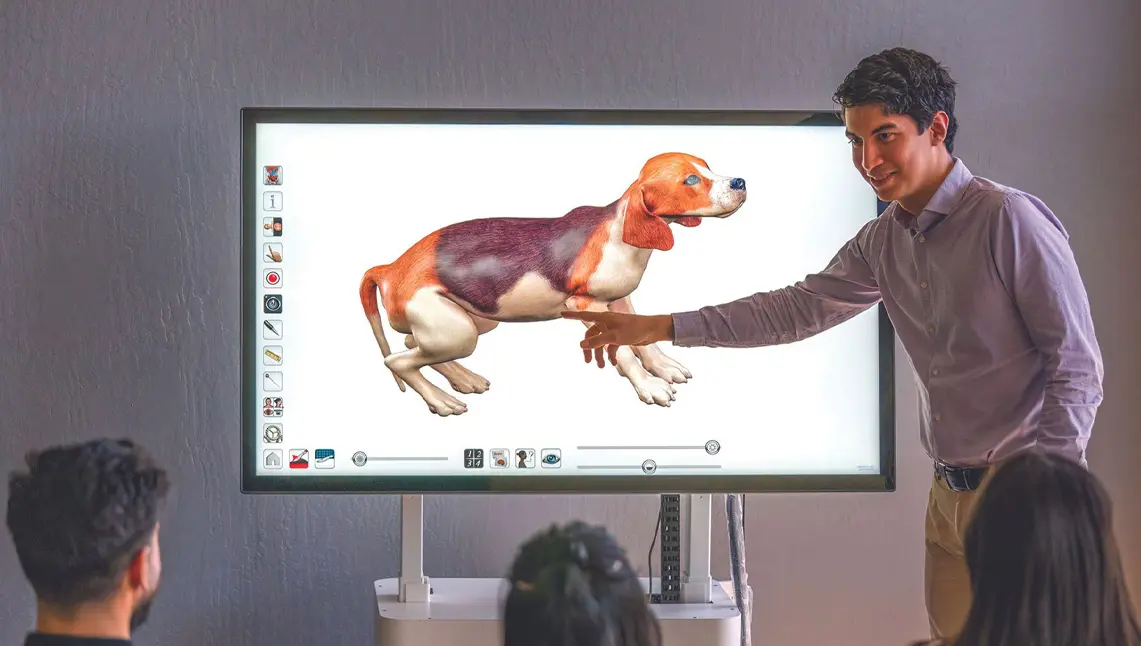
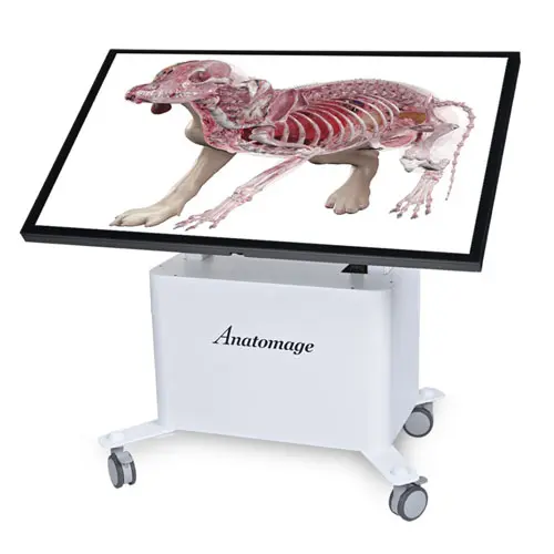
The Anatomage Table Vet is a digital animal dissection platform that offers comprehensive anatomy resources in 3D for veterinary medicine, animal science, and zoology. With its digital real-tissue animal cadavers and extensive pathology library, the Table Vet aims to improve animal anatomy learning outcomes while setting technological standards for veterinary institutes.
Anatomage Table Vet
Anatomage Table Vet:
Bring Realism to Veterinary Science

The future of veterinary science is here

Mobility.
Visually compare animal anatomy with humans through a diverse case library consisting of comparative study cases with synchronized dissections of multiple cases. Students can learn to identify anatomical differences between human and animal bodies.
Visually compare animal anatomy with humans through a diverse case library consisting of comparative study cases with synchronized dissections of multiple cases. Students can learn to identify anatomical differences between human and animal bodies.
3D Realism.
Bring realism into veterinary education by providing an absolutely explicit 3D visualization of animal anatomy. The platform features 3D animal cadavers – which include the world’s most detailed canine cadaver – that assist in animal dissection.
Anatomical Variations.
Introduce a high-quality approach to animal learning using digital veterinary models. Unlike physical animal cadavers, the Anatomage Table Vet’s bodies are reconstructed from actual animal anatomy data allowing for correct visualization during dissection.
World’s Most Accurate Canine Cadaver
The Anatomage Dog is the first highly detailed dog anatomy atlas that comprehensively features internal organs, including vascular systems and muscular-skeletal structures. Originating from real dog data, Anatomage Dog exhibits the highest level of animal anatomy accuracy. All of its volumetric 3D and individual structures are segmented, so users can peel off structures layer-by-layer to reveal inner details. Individual structures are also fully annotated.
1,185 Structures.
Our Anatomage Dog is the most detailed canine cadaver featuring highly segmented and annotated structures. Vascular and musculoskeletal structures can be visualized in clear 3D detail, greatly enhancing the study of canine anatomy.
Animal Dissection.
Deepen your understanding of gross anatomy by interactively dissecting the canine body. Visualize internal animal anatomy with tools that allow rotation, multiple cuts, and instant undo. Easily memorize canine anatomy terminology with annotated structures.
Assessments.
Effortlessly create quizzing materials using the Anatomage Table Vet’s 3D animal anatomy content. Conduct interactive and collaborative assessments with Quiz Mode, and easily monitor student performance with report exporting features.
Quick Facts
Explore these quick facts about the Anatomage Table Vet.

Anatomy & Physiology
- Real-tissue Animal Cadavers
- 285 Real Animal Scans
- Histology Slides
- Real Cadaveric Prosection Scans
- Functional Anatomy
Features & Functionalities
- Lifesize Full Body Display
- Adjustable Display Angle
- Video Output Capability
- Upload & Render Medical Scans
- PACS Compatibility
- Ultra High Quality for CT & MRI
- Anatomical Radiology Curriculum
- High Resolution Regional Anatomy
Discover The Applications
Animal Dissection
The Anatomage Table Vet promotes ethical animal dissection practices, eliminating the risk of harmful chemical exposure.
Veterinary Training
The tool enhances hands-on learning for veterinary trainers with its 3D animal dissection capabilities, real-life cases, and comprehensive animal anatomy details.
Animal Science & Pathology
The Anatomage Table Vet offers CT/MRI scans that display both normal and abnormal animal anatomy across various species, providing invaluable insights into animal pathology.
Lecture Aids
The Anatomage Table Vet can connect to projectors, displaying 3D visuals of animal anatomy and pathology. It also fosters classroom collaboration by enabling content sharing through screenshots.
