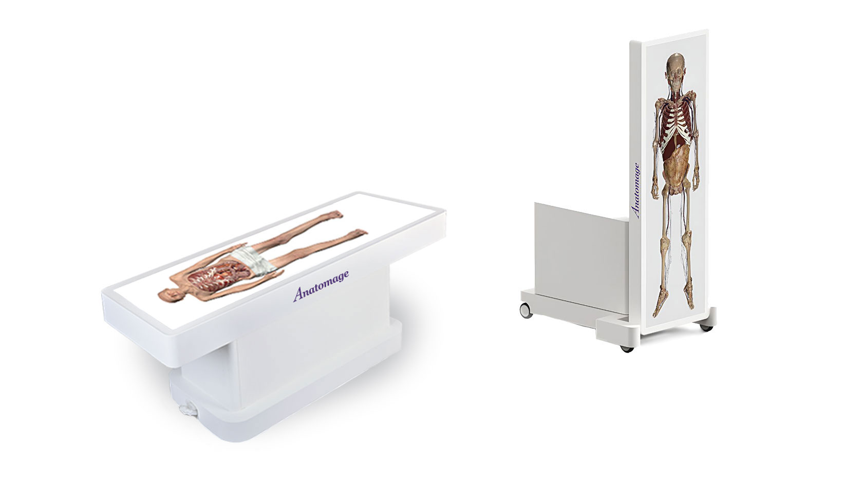
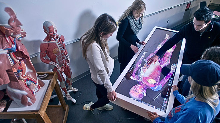
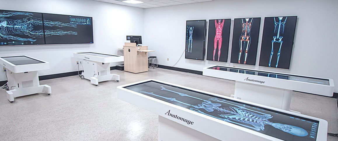
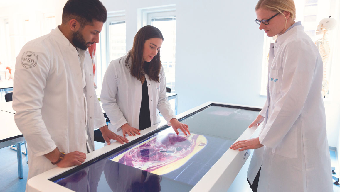
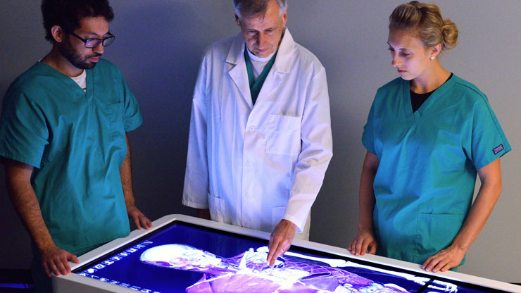
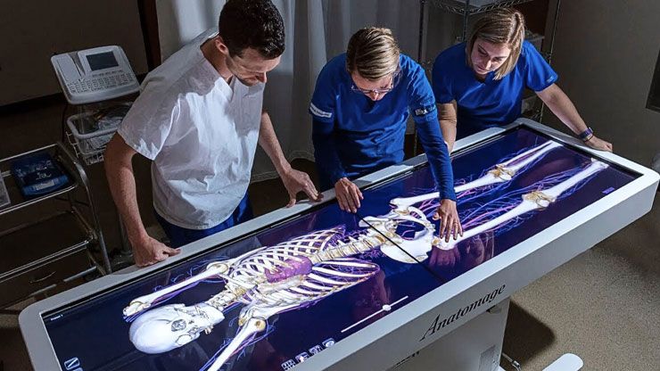
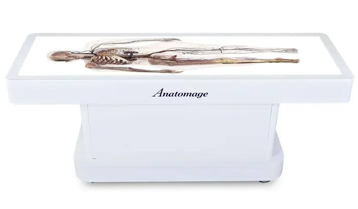
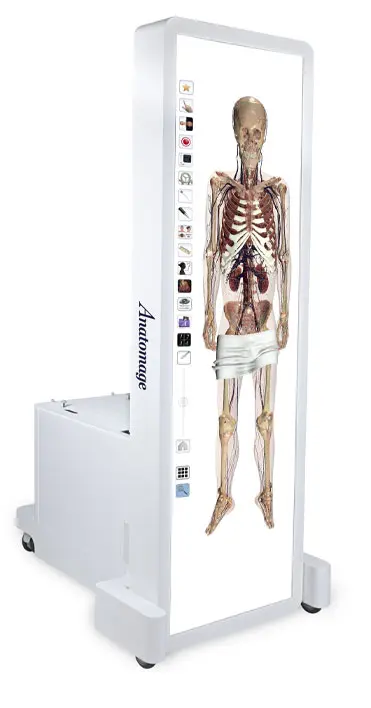
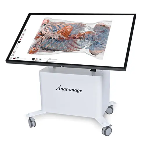
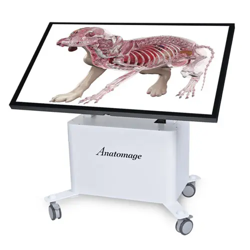
Journey through life in 3D

Our bodies have stories to tell. And Anatomage Table 10 brings those stories to life. Furthering the Anatomage Table’s legacy of digitizing real anatomy, Table 10 opens a new dimension into exploring anatomy in a way that was previously impossible. From simulating the stages of childbirth to tracing the effects of disease on an aging body, Table 10 provides a window into life–a scientific journey into what anatomy can teach us. The Anatomage Table, now with the enhancement of Table 10, is more than a learning platform; it deepens clinical care through anatomical insights.

Not a Table user?

See it in action

High-resolution

Table 10’s high-resolution rendering brings our Anatomage Bodies into sharp focus, displaying even the smallest details down to 0.2 mm in clear, photorealistic detail. This creates an immersive learning experience where every anatomical detail can be studied.

Diverse case studies

Our Case Studies library has expanded to include additional histology, sea animals, and botany. This allows students to explore life beyond human anatomy, revealing shared anatomical similarities among species.

Learning to experience

Table 10 provides an environment for medical students and trainees to learn and practice clinical scenarios. New features, including intuitive instruction tools and interactive, diagnostic techniques, are available to foster a practical learning space.

A new geriatric cadaver

Discover the marks diseases leave on an aging body. The Table 10 software introduces a new male cadaver, called Hans, reconstructed from an actual 70-year-old patient who passed away from lung cancer. The new Anatomage Body offers insights into a range of pathologies, including lung cancer, and tumors in the thoracic wall, pancreas, and liver. Utilizing Table 10, students and professionals can investigate the anatomical manifestations of both aging and diseases via high-resolution 3D visualizations.

  • Real anatomy
  • High-resolution at 0.2 mm to 1.0 mm
  • Ideal for geriatrics, oncology

Childbirth simulation

The Birth Simulation, a new feature on Table 10, presents the first-ever fully 3D interactive simulation of childbirth, featuring authentic human anatomy. This advanced simulator offers extreme visual clarity into the various stages of labor, including the cervical dilation, infant rotation and head movements, and release of the placenta. Medical professionals and students can remove anatomical parts to explore the anatomical transformations occurring throughout the birthing process.

  • Interactive interface
  • Adjustable speed
  • Ideal for obstetrics, maternal education and care

Arrhythmias

Our upgraded Cardiology simulation now showcases a collection of arrhythmias, enabling students to compare a normal heartbeat with irregular rhythms such as atrial fibrillation, ventricular fibrillation, and various heart blocks. Clinical professionals can identify relevant clinical anatomy at different heart rates and investigate what they mean for their patients’ cardiovascular well-being. 

  • Cardiac activity can be tracked on an ECG
  • A-Fib, V-Fib, and 1st, 2nd, and 3rd degree heart blocks
  • Ideal for cardiovascular education and training

Vascular Grow

As part of our Blood Flow tool, the Vascular Grow feature maps out the blood vessels in the cadaver, giving medical students a thorough view of how the heart and blood vessels are connected. Doctors can use this tool to see where important arteries split and veins come together. It’s also helpful for explaining to heart patients how their blood vessels work and where to place catheters safely.

  • Interactive view of the blood flow
  • Visualize arterial bifurcation and venous convergence
  • Identify vascular anomalies or conditions

Ultrasound Viewer

The Ultrasound Viewer offers real-time, cross-sectional imaging that mimics actual ultrasound technology, revealing the internal structures of a cadaver in supreme quality. Suitable for both educational and professional settings, this tool allows learners to place a probe on the simulated body, generating corresponding sectional views. As an ideal resource for sonography training, the Ultrasound Viewer not only enhances understanding of human anatomy but also teaches the impact of probe rotation and angle on the resulting images.

  • Ultrasound Fan View
  • Real-time, cross-sectional images
  • Ideal for sonography, radiology

Point-to-point Dissection

Point-to-point dissection is a step-by-step procedure that helps students easily visualize anatomical structures and diseases. Students initiate the process by marking an entry point, then proceed to dissect through a connected point. This method aids students in understanding the spatial interrelationships between different anatomical elements and provides a comprehensive perspective for examining tumors from multiple angles.

  • Explore the spatial relationships
  • Practice customized surgical procedures
  • Ideal for surgical planning, sport injuries inspection

Clinical-Based Learning Tools

We simplify learning. Our tools enable students to quickly identify anatomical structures and distinguish adjacent structures through various colors. In addition, students can now customize their Anatomage Table’s navigation. These user-friendly tools not only enhance student engagement with the subject but also create a more effective learning experience.

  • Color tools
  • Personalized navigation
  • Ideal for group sessions, autonomous learning
Contact Anatomage for details