

Every year, 1 in 4 people die from heart disease. With cardiovascular disease continuing to be the leading death cause in the United States – and the rest of the world – cardiac care inspires innovation in technology development. Many innovations are made to better the understanding of the heart.
Amidst the advancements, cardiac simulation plays a special role. Let’s take a look at how simulation can simplify the anatomical and physiological complexity of the heart.
Simulation of regular cardiac conduction systems
As one of the most complex organs, the heart’s internal anatomical and physiological activities operate through its electrical mechanism. With that said, being able to conceptualize the heart’s electrical system is critical to understanding cardiac pathophysiology. And simulation allows for such conceptualization.
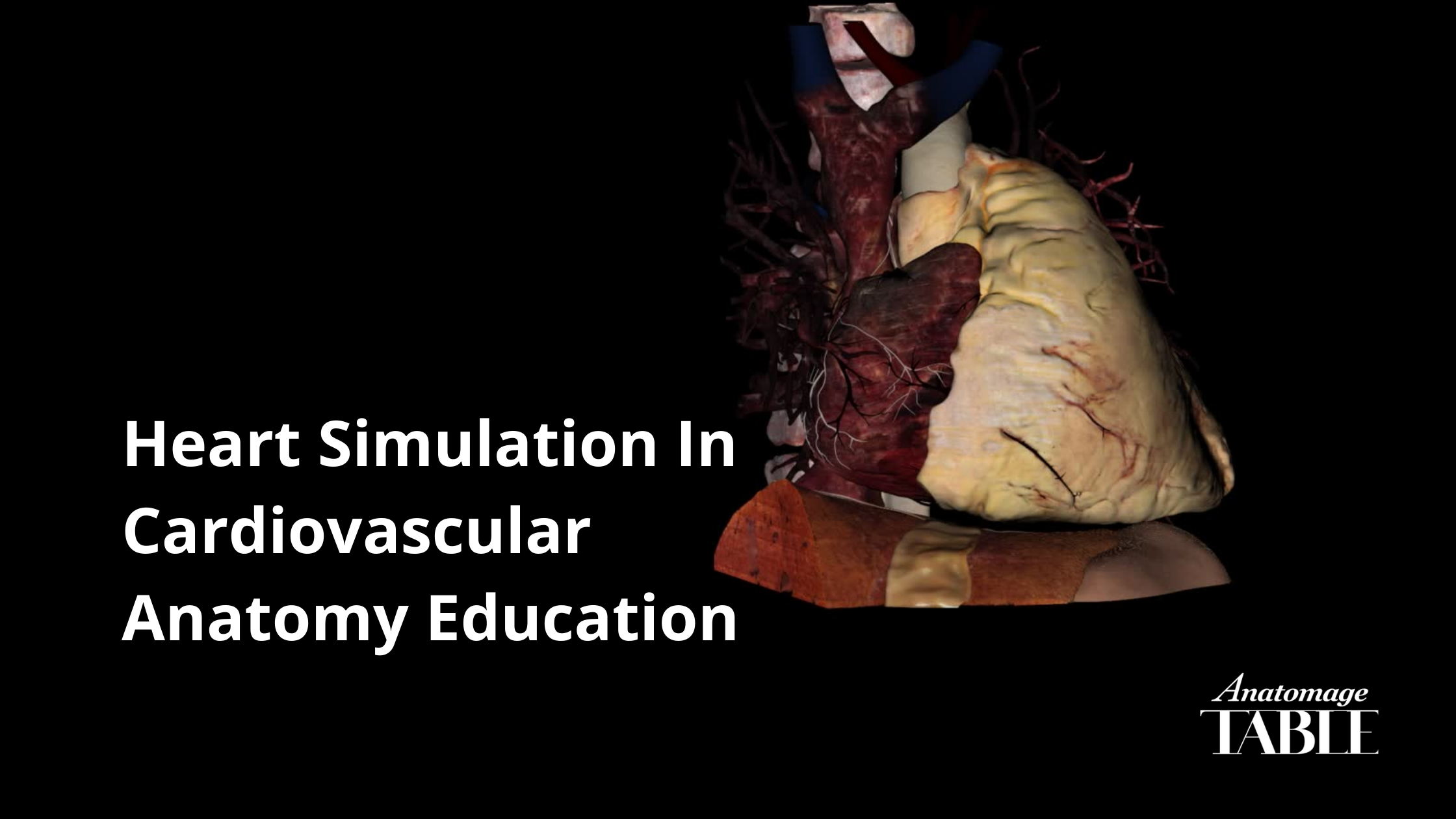
The video depicts the heart motion simulated using actual heart CT images.
The sinoatrial (SA) node – which is located in the right atrium of the heart – is responsible for generating the electrical stimulus. In the video, the SA node is noted in yellow color. Once the node sends out the electrical signals through the muscle cells, the upper atria begin to contract. Noted by the purple circle, the atria then pump blood into the ventricles. When the electrical impulse travels to the atrioventricular (VA) node, it will be shown down to give time for the ventricles to receive the blood. Once the ventricles begin to contract, the right ventricle carries blood to the lung and the left brings blood to the rest of the body. Then the SA node sends another signal to repeat the cycle. Normally, a heart beats 60 to 100 beats per minute.
It’s important to visualize the normal cardiac electrical system to conceptualize how the heart works properly. This serves as a strong foundation to investigate and study abnormal cardiac rhythm conditions – like heart arrhythmias. In the Anatomage Table, the heart motion tool can provide an accurate representation of how the heart works.
Simulation of spatial relations of the heart anatomy
Another benefit of heart simulation is that it allows for the visualization of spatial anatomical relationships. Spatial relations of ventricular structures can’t be taught by static images or textbooks. The fundamentals of spatial relationships in cardiovascular anatomy require visual perception.
A report published by Yenepoya Medical College surveying 145 medical students demonstrates that the simulation-based teaching of cardiovascular physiology enhances medical learning and applications.
The college set up a learning environment consisting of four stations:
- The first station included the Anatomage Table
- The second station had an ultrasound simulator that allowed students to visualize cardiac hemodynamic
- The third station had a human patient simulator in which students could learn about the electrical conduction system of the heart through detecting the abnormalities of cardiac rhythm in ECG
- The last station also included a patient simulator that allowed students to perform a clinical examination of the cardiovascular system.

Image from ijpp.com
After completing the learning module involving interacting with these four stations, 84.3% of the students strongly agreed that the use of the Anatomage Table improved their understanding of cardiovascular anatomy. For instance, students could perform layer-by-layer dissection and photo-realistically visualized the spatial relations of internal anatomy structures in various viewing 3D planes on the Table.
Providing 3D visualization for the cardiac cycle and spatial relations of cardiac structures are just a few of what cardiac simulation technology can bring to cardiovascular anatomy teaching.
Learn more about Anatomage’s 3D modeling and simulation here.
Other content