
Case Study: Life University
Case Study:
Life University
Modernizing Education: How Life University Reimagines Chiropractic Anatomy Education with the Anatomage Table
Replacing Traditional Cadaver Dissection with Clinically Relevant 3D Technology for Future Chiropractors
Back in 2014, Life University, a leader in chiropractic education, faced challenges with their traditional cadaver laboratory. Issues like escalating costs and updates to chemical handling regulations in their cadaver facilities complicated operations. More fundamentally, Life’s faculty recognized a disconnect between their educational methods and clinical reality: Chiropractors would never perform surgical dissection in practice, but their students were spending a significant amount of time developing dissection skills they would never use.
So, the university transformed their anatomy instruction with the Anatomage Table, a virtual dissection solution that aligns with the chiropractic scope of practice while providing superior learning outcomes, all without the limitations of traditional cadaver labs.
Challenge
For years, Life University’s Doctor of Chiropractic program relied on cadaver labs for gross anatomy instruction that were costly, time-consuming, and challenging to manage. The financial burden of maintaining a traditional cadaver laboratory proved substantial, including acquisition costs, chemical preservation systems, and specialized ventilation. Additionally, the state’s changing regulations around chemical handling and storage added complexity and expense to an already resource-intensive operation. These compounding issues raised concerns about the long-term sustainability of a cadaver-based approach.
More importantly, traditional dissection created barriers that hindered rather than enhanced learning for Life University’s chiropractic students. They were developing motor skills for dissection procedures they would never perform professionally, taking valuable time and focus away from actually understanding anatomy. “We’re training chiropractors,” said Dr. Eric Partin, an assistant professor at Life University. “We don’t need to cut into cadavers… You will never do that as a chiropractor.”
The cadaver format also limited study flexibility and accessibility. Students could only study cadaveric anatomy during scheduled lab times, restricting their ability to review material at their own pace. Life’s faculty recognized that these barriers prevented students from fully engaging with anatomy and from applying their understanding to chiropractic practice.
At the same time, the university sought to modernize its teaching methods to better align with modern students: learners who thrive with interactive technology. The faculty aimed to create an anatomy program that was not only comprehensive and accurate, but also accessible, engaging, and relevant to the clinical realities of chiropractic.
Solution
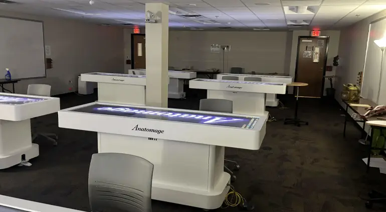
To address these challenges, the leadership at Life University made the pivotal decision to transition to fully digital anatomy instruction. Initially introducing eight Anatomage Tables across its anatomy courses in 2015, the university currently has 19 Tables at their state-of-the-art Virtual Anatomy Lab.
To maximize the Table’s educational value while addressing chiropractic-specific learning objectives, Dr. Partin developed a comprehensive implementation strategy by creating customized preset materials aligned with laboratory lists. Class sessions begin with instructor-led demonstrations displayed on wall-mounted screens, allowing students to follow along as faculty navigate through anatomical structures and relationships.
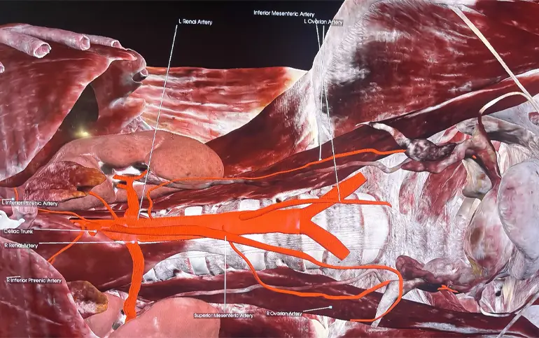
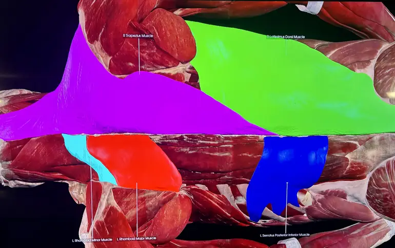
Then, using the Tables, students transition to small-group work, where they independently explore the same preset materials demonstrated. They can conduct full-body virtual dissections, explore cross-sectional anatomy, and analyze 3D structures with precision and clarity. The ability to remove and restore layers instantly, highlight specific systems, and view anatomical structures from multiple perspectives allows for an unparalleled level of interactivity and comprehension.
On top of that, the peer-assisted learning approach that comes with small groups allows students to work collaboratively while building confidence in their anatomical knowledge. Instructors also utilize the self-test functionality, encouraging students to identify knowledge gaps and focus study efforts efficiently rather than reviewing already-mastered content. Students even have access to the Tables during open lab times, enabling flexible study scheduling that accommodates their demanding academic programs.
The Anatomage Table is a wonderful alternative because cadaver labs are extremely expensive. And the Tables are so much better than just using anatomical models.
Dr. Eric Partin, Assistant Professor, Life University
Results
Since adopting the Anatomage Table, Life University has seen fundamental transformations to their chiropractic anatomy education across multiple dimensions. Without the need for cadavers or chemical storage, the university has significantly reduced recurring operational costs while creating a safer and more sustainable lab environment. The virtual lab setup has also made the anatomy courses more accessible to all students, particularly given that their current cohorts grew up with touchscreen devices and have native familiarity with the interface.
Engagement-wise, students have demonstrated greater willingness to manipulate and explore anatomical structures, and the technology’s intuitive nature means students spend minimal time learning to use the system and maximum time studying anatomical content. “They like using the Table as opposed to using cadavers,” shared Dr. Partin. “They have the information that I went over in class there so they can study during their open lab times, having some flexibility to review on their own time.”
The implementation of the Anatomage Table has also contributed to improvement in academic outcomes. In a study performed after the Tables’ introduction at Life University, faculty found that students learning and testing with the Table performed at or above the level of peers who previously studied with cadavers or anatomical models. Faculty members attributed this success to the Table’s clear visual detail and the hands-on engagement, which helped students focus more on understanding spatial relationships between structures rather than the mechanics of dissection.
Beyond the classroom, the Anatomage Tables have become a powerful recruitment tool and a signature program differentiator. Prospective students and families visiting Life University cite the Virtual Anatomy Lab as “one of the highlights of the tour,” with the Tables generating significant interest among applicants. The university’s position as home to the one of the world’s largest standalone Anatomage labs helps attract students interested in learning through modern, technology-enhanced methods.
Life’s faculty are continuing to redefine their curricula and integrate more Anatomage Table content in their classrooms, like incorporating the Table’s built-in CT and MRI images and clinical case studies. The university has already started introducing the Table’s capabilities to their growing undergraduate program and sees the Table as an evolving cornerstone of their teaching. As the university’s programs continue to expand, faculty envision the Anatomage Table playing an even greater role in shaping how future chiropractors learn anatomy and clinical reasoning. By embracing virtual 3D anatomy, Life University has not only eliminated the costs and complications of traditional cadaver labs, but also set a new standard for how technology can elevate chiropractic education.