
Case Study: Orange County Medical Examiner
Case Study:
Orange County Medical Examiner
Photo provided by Orange County District 9 Medical Examiner’s Office
Revolutionizing Forensic Investigation: How a Medical Examiner's Office Enhanced Case Analysis with the Anatomage Table
Transforming Post-Mortem CT Analysis with 3D Visualization Technology
When the District 9 Medical Examiner’s Office in Orange County, Florida acquired a CT machine in their new facility, Chief Medical Examiner Dr. Joshua Stephany faced a significant challenge: how to maximize the diagnostic potential of 2D CT scans without extensive radiology training. They needed an innovative solution that could seamlessly integrate with their existing workflow while providing clearer, more comprehensive case analysis capabilities.
To complement the office’s use of CT imaging, Dr. Stephany and his team have integrated the Anatomage Table Clinical, which has become an indispensable tool in postmortem examination, trauma assessment, and forensic education. As the only medical examiner’s office in the country using this technology, Dr. Stephany has pioneered new workflows that combine visual clarity with digital efficiency.
Challenge
The medical examiner’s office encountered significant obstacles in maximizing their forensic investigation capabilities. With increasing caseloads and a national shortage of forensic pathologists, medical examiner’s offices across the country have turned to CT machines to aid with postmortem examinations.; however, traditional 2D CT analysis requires extensive radiology expertise that most medical examiners don’t possess, limiting the technology’s diagnostic potential. “We did not want to relearn two dimensional CTs,” explained Dr. Stephany, highlighting the need for more intuitive visualization tools that could integrate seamlessly with their existing workflow.
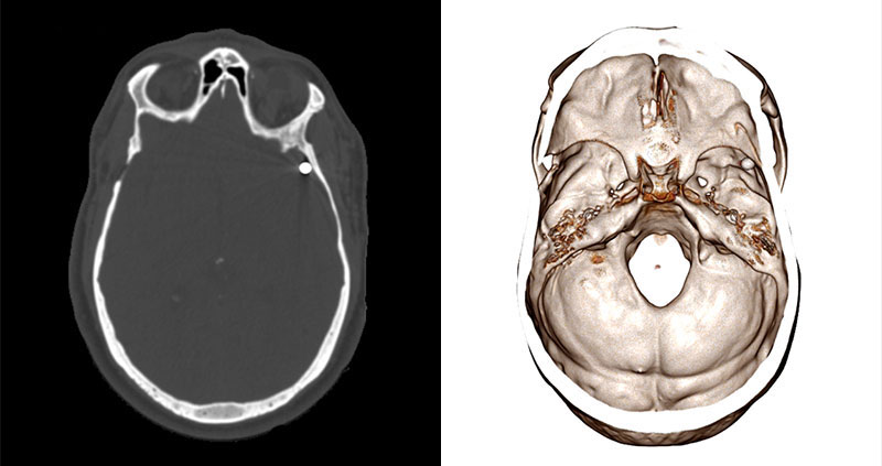
Anatomage Case Library: Gun Shot Wound. CT Scan (left), Anatomage Volumetric Rendering (right).
Complex trauma cases, like with multiple gunshot wounds, could also present particular challenges, requiring time-consuming X-ray procedures with repeated plate movements and manual synchronization. Locating projectiles and understanding their trajectories in traditional 2D imaging was labor-intensive and not as precise, often leaving doctors with incomplete spatial understanding of complex trauma patterns. With the need for rapid case resolution, the office required a supplemental tool that could streamline their investigative process while maintaining diagnostic accuracy.
Solution
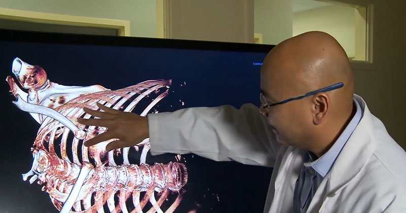
Photo provided by Orange County District 9 Medical Examiner’s Office
In 2020, Dr. Stephany’s office implemented the Anatomage Table Clinical as a comprehensive 3D visualization solution, strategically integrating it into their processes to address these challenges. The Table seamlessly uploads postmortem CT data through DICOM integration, enabling immediate and interactive 3D reconstructions of cases for detailed analysis — all without requiring extensive radiology training.
Dr. Stephany’s team uses the technology flexibly throughout their investigative workflow, applying it both pre-autopsy for case planning and post-autopsy for documentation. Approximately 80% of their usage focuses on obtaining high-quality images for case files and potential court presentations, while the remaining time is dedicated to pre-autopsy case assessment and planning. In certain cases, the team also utilizes the Anatomage Table Clinical to triage cases before autopsy — particularly in elderly individuals where coronary disease may be visually confirmed in advance. As Dr. Stephany explained, “The Anatomage Table is really good on trauma and broken bones and projectiles… And we’re getting better at diagnosing [conditions like] cardiac tamponade or aortic dissection prior to autopsy.”
The office has established comprehensive protocols for Anatomage Table Clinical usage, ensuring all homicides, decomposed remains, infant cases, and gunshot wounds receive CT scanning with subsequent 3D analysis capabilities. This systematic approach allows for detailed trauma assessment while serving as a powerful communication tool for law enforcement briefings, attorney consultations, and family discussions. Plus, the Anatomage Table Clinical’s mobility within the facility enables the team to move it between rooms as needed while keeping it outside the morgue environment to maintain optimal cleanliness.
I say to other medical examiners: ‘If you’re going to get a CT, the Anatomage Table is a great supplemental tool.’ It makes everything we do with CTs that much better and that much easier.
Dr. Joshua Stephany, Chief Medical Examiner at District 9 Medical Examiner’s Office
Results
The Anatomage Table Clinical has delivered transformative results across multiple aspects of forensic investigation, saving time and fundamentally changing how the office approaches complex cases. Cases with multiple projectiles or fractured bones now require significantly less time for location and analysis, with doctors able to quickly rotate 3D models to understand spatial relationships.
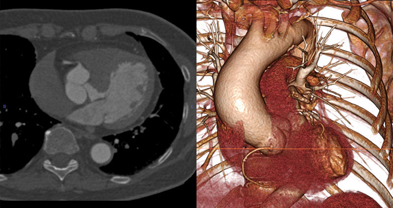
Anatomage Case Library: Stable Thoracic Aneurysm. CT Scan (left), Anatomage Volumetric Rendering (right).
The technology has also enhanced the office’s diagnostic capabilities, improving their ability to diagnose conditions like coronary disease and subdural hemorrhages without the need for full autopsy procedures. This advancement enables more efficient case resolution while providing families with clearer explanations supported by visual evidence. High-resolution 3D images also serve as compelling visual evidence for court presentations, with saved screenshots and JPEGs readily available for legal proceedings.
From a professional development perspective, new medical examiners and staff quickly adapt to the technology, particularly recent graduates who are already familiar with 3D anatomy visualization from their educational programs. The Anatomage Table Clinical has also expanded the office’s investigative capabilities, facilitating identification of unknown remains through ante-mortem to post-mortem comparisons and providing detailed bone fracture analysis that would be difficult to visualize during traditional autopsy procedures.
Dr. Stephany describes the overall impact succinctly: “It’s a game changer. I used to tell people what they see on TV isn’t real, but now with the Table, I can rotate the body, remove tissue, and it has become real.”
By positioning the Anatomage Table Clinical as a central component of their forensic workflow, this Orange County medical examiner’s office has enhanced their diagnostic capabilities and established a new standard for modern forensic investigation — demonstrating how other offices can achieve similar advances in accuracy and efficiency.