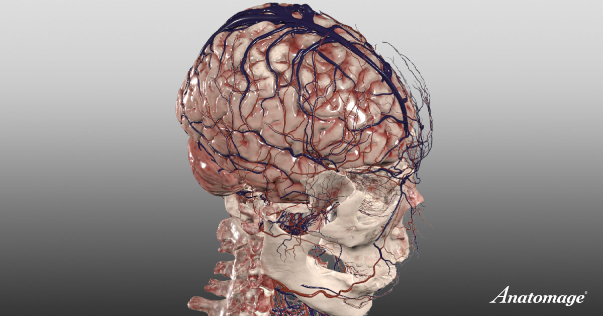
In human anatomy, the brain is recognized as the most complex organ, consisting of approximately 86 billion neurons. Each neuron forms extensive networks of synaptic connections, which are foundational to cognition, memory, and autonomic function. High-resolution brain models and 3D anatomy visualization tools have made it easier to study these connections in clinical and educational settings.
According to a landmark study published in the Journal of Comparative Neurology, the cerebral cortex alone contains around 16 billion neurons, emphasizing the intricate structure and function of the human nervous system.
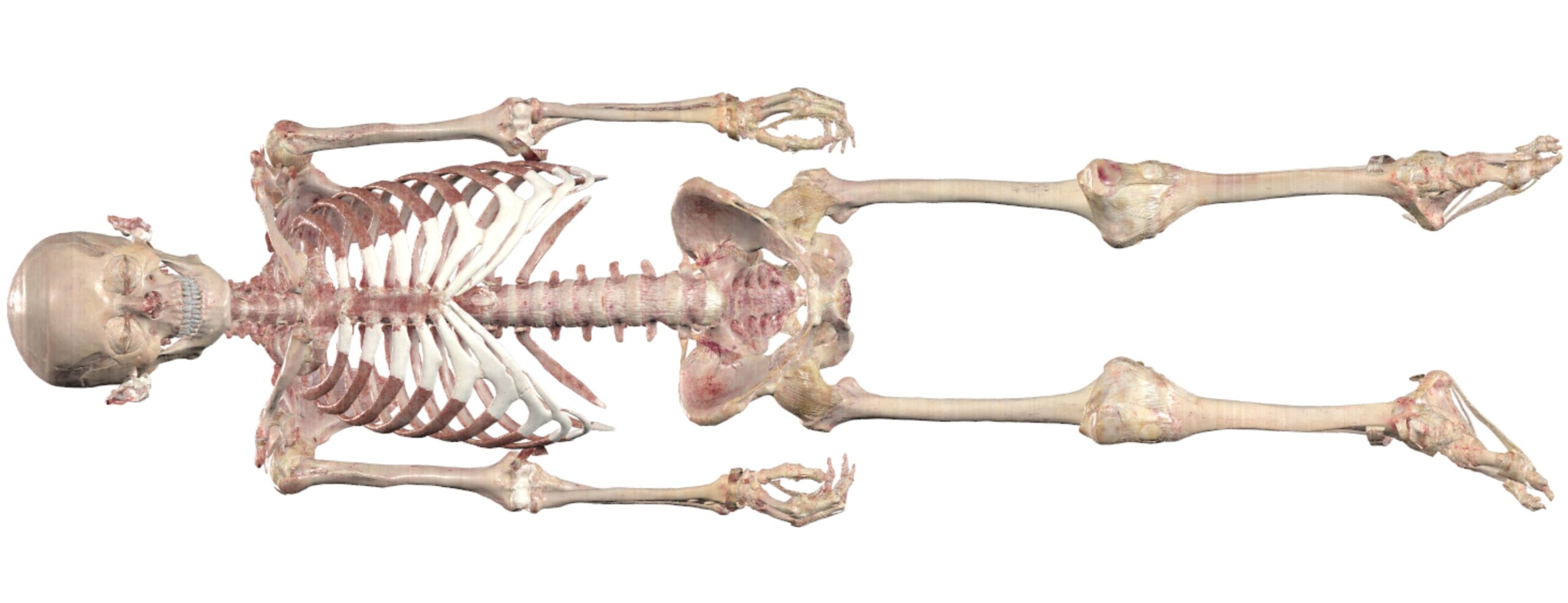
Fact 2: Foundational Anatomy of the Adult Human Skeleton
A core concept in skeletal anatomy is that the adult human skeleton comprises 206 bones, reduced from around 270 in infancy due to bone fusion during development. These bones provide the body with structure, protect internal organs, and support movement through complex joint systems.
In clinical anatomy and anatomical modeling, understanding bone morphology is essential for diagnosing musculoskeletal conditions and planning surgical interventions. Bone models are also widely used in medical education for hands-on exploration of structural relationships and articulations within the skeletal system.
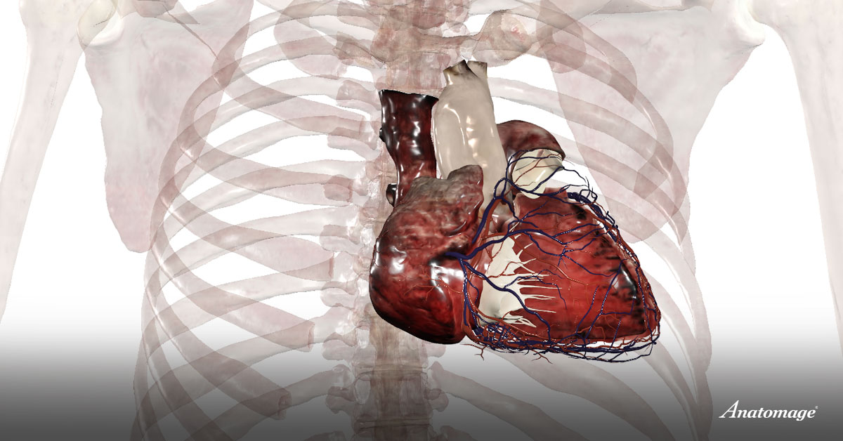
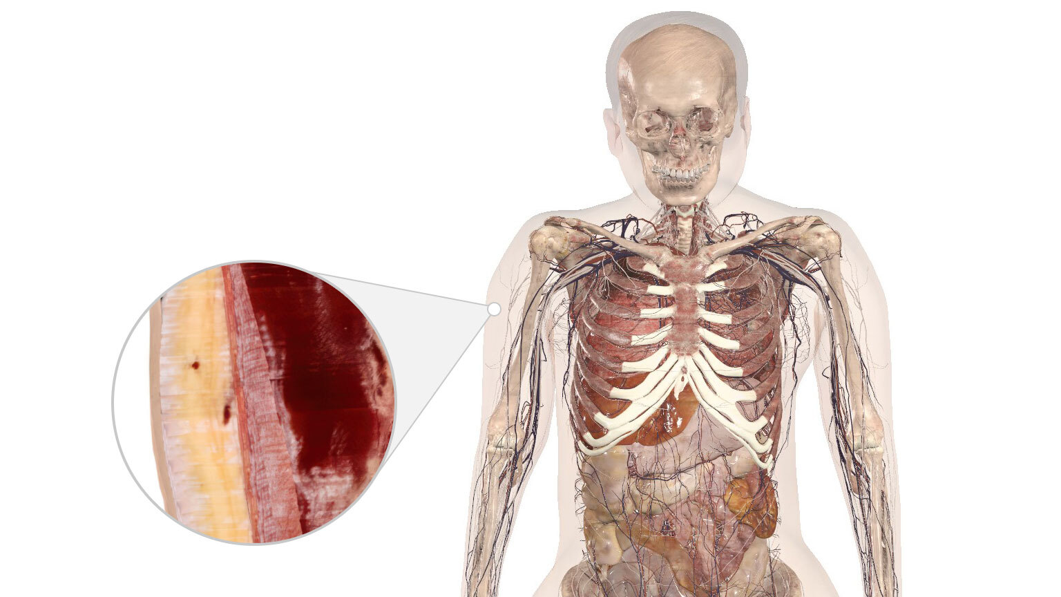
Fact 3: Functional Capacity of the Human Heart in Systemic Circulation
Cardiovascular anatomy highlights the heart as a muscular organ that propels roughly 7,500 liters of blood through the circulatory system each day. Detailed heart models help illustrate the four-chambered structure, valves, and conduction pathways responsible for maintaining rhythmic and unidirectional blood flow. Advanced tools in clinical anatomy allow for the simulation of heart conditions and surgical planning, improving both patient care and medical education.
According to the American Heart Association, the average adult heart beats around 100,000 times per day to maintain continuous systemic and pulmonary circulation.
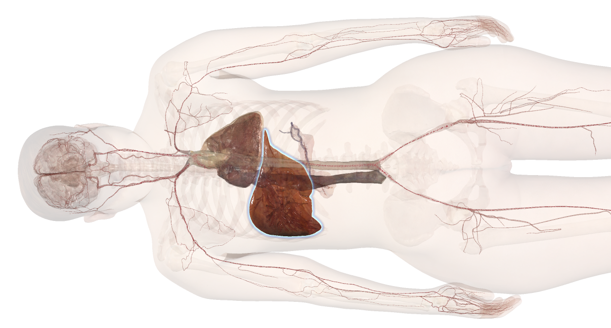
Fact 4: The Liver as a Central Organ in Human Anatomy and Physiology
As the largest internal organ in human anatomy, the liver is involved in over 500 essential physiological processes, including detoxification, protein synthesis, and bile production. Its anatomy is defined by distinct lobes, a dual blood supply, and a complex biliary system.
Understanding liver anatomy is fundamental in clinical education, particularly when studying digestive function, metabolic regulation, and systemic circulation. According to Johns Hopkins Medicine, the liver’s extensive functionality makes it one of the most vital and versatile organs in the human body.
Fact 5: How the Skin Protects and Connects
The skin is the largest organ in human anatomy, covering the entire body and playing a vital role in protection and regulation. It consists of three main layers: the epidermis, dermis, and subcutaneous tissue—each contributing to functions such as temperature control, immune defense, and sensory perception. In addition to acting as a barrier against environmental threats, the skin supports the production of vitamin D and helps retain essential fluids.
According to the U.S. National Library of Medicine, the skin spans about 1.5 to 2 square meters in surface area and accounts for roughly 16% of an adult’s body weight.
References:
Azevedo FA et al., Journal of Comparative Neurology (2009)
Gray’s Anatomy, 41st Edition, Elsevier
American Heart Association – How the Heart Works
Johns Hopkins Medicine – Liver Anatomy and Function
MedlinePlus (U.S. National Library of Medicine) – Skin Overview
Anatomage Table
For explore more human anatomy, check out the Anatomage Table.