Our case studies explore how the Anatomage Table has been integrated into learning and training programs across hospitals, clinics, and institutions in the US and around the world. The Table’s impact has led to significant qualitative and quantitative improvements for both students and medical professionals. Explore the case studies below to discover how various institutions have seamlessly integrated the Anatomage Table into their curricula.
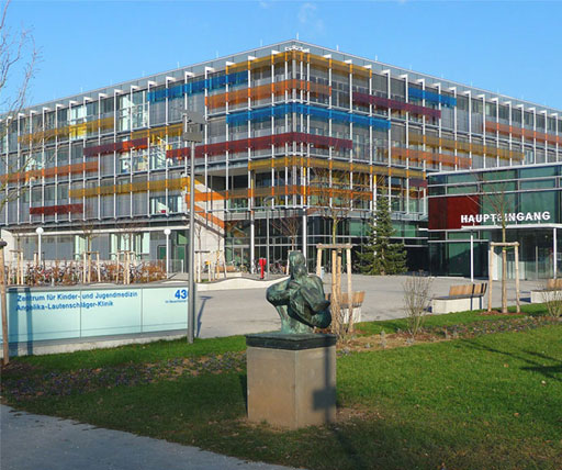
University of Heidelberg Medical School
Anatomage Table Increased Student Scores by Up to 27%
In this case study, discover the results of an assessment conducted by researchers from the University of Heidelberg, the German Cancer Research Center, and the Karlsruhe Institute of Technology. They collaborated to evaluate the benefits of Anatomage Tables and cadaver CT scans on student learning of radiologic anatomy.
Anatomage Table Proven Effective for Anatomy Education Compared to Cadaver-Based Teaching
Faculty members at Life University conducted an evaluation of lecture and laboratory scores of first-year chiropractic anatomy students. Their goal was to determine if students learning with the Anatomage Table could achieve similar learning objectives as those using traditional models or cadavers.
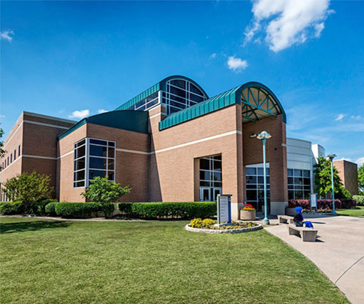
Anatomage Table Offers Great Benefits for Anatomy Courses in Community College Setting
Tarrant County College acquired three Anatomage Tables for their various campus locations to enhance their Physical Therapy Assistant, Anatomy & Physiology, and Emergency Medical Technician programs. Additionally, student perceptions of the Table were explored through a qualitative research study focusing on radiographic positioning courses.
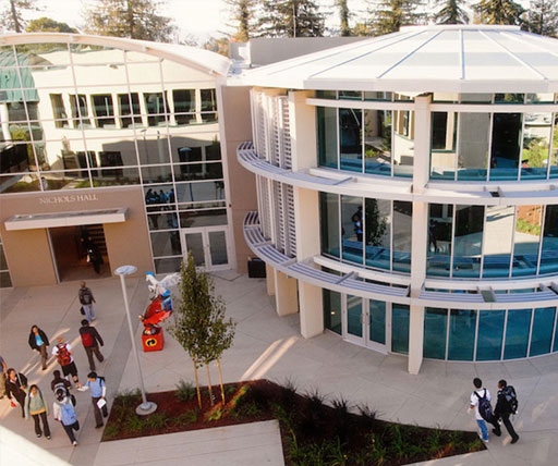
Anatomage Table Receives Excellent Reviews from Stanford Program Students
Students in the Clinical Anatomy Summer Program (CASP) at Stanford University interact hands-on with human anatomy while learning foundational medical skills. CASP exposes high school students to dissection practices, surgical procedures, and biomedical technologies.
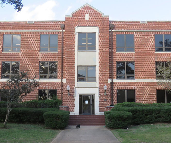
Anatomage Table Allows High School Students to Access Virtual Human Cadavers
Explore how The Harker School, the first private high school to acquire the Anatomage Table, revolutionized its anatomy education. With no access to traditional cadavers, students thrived using digital cadavers and virtual dissection tools, significantly enhancing their learning experience. Focused on AP Biology, Human Anatomy, and Physiology courses, this innovative approach fostered student engagement, critical thinking, and mastery of anatomy terminology and dissection.
Promoting Spatial & Conceptual Learning of Anatomy in Biology Courses
Prairie View A&M University’s Biology Department is transforming science courses with the Anatomage Table. With classes of 32 to 64 students and 500 biology majors, the Table is a key resource. Used in over five courses, it enhances learning in gross anatomy, physiology, biology, and radiology technician training with real-time X-ray visualization. Students benefit from virtual dissection and advanced anatomy education, responding enthusiastically to this innovative tool.
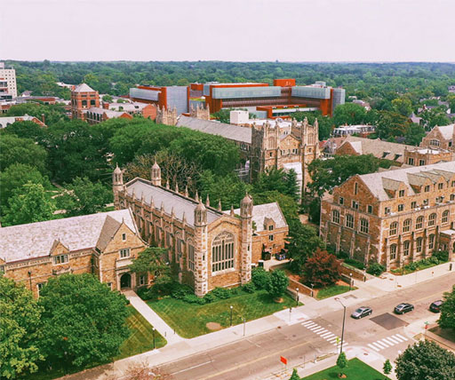
Nursing Program Successfully Adopts Anatomage Table
The University of Michigan’s undergraduate nursing program has successfully integrated the Anatomage Table into its curriculum, revolutionizing anatomy and physiology courses with hands-on, interactive learning. Discover how Anatomage Table enhances student engagement and understanding in our detailed case study.
Anatomage Table Increases Exam Scores and GPA for Imaging Science Program
Discover how the University of Nebraska Medical Center enhanced their medical imaging courses with the Anatomage Table, leading to improved exam scores in gastroenterology (GI) courses. Learn about the integration process, benefits, and positive outcomes in our comprehensive case study.
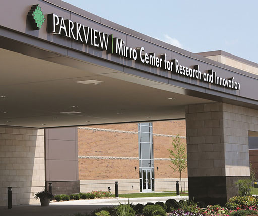
Clinical Education & Surgical Planning with Anatomage Table in Simulation Labs
The Parkview Mirro Advanced Medical Simulation Lab utilizes the Anatomage Table across various disciplines and health services. It educates healthcare providers, including surgeons, physical therapists, and occupational therapists. The Table’s capability to view and virtually dissect real patient 3D images makes it invaluable for endovascular procedures and surgical planning. Additionally, it serves as a powerful patient education tool, enhancing doctor-patient understanding and boosting patient satisfaction.
Read the full case study to learn more about the impact of the Anatomage Table.
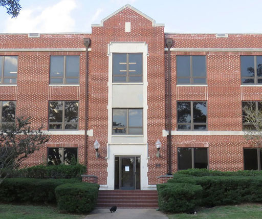
Winthrop University Uses Anatomage Technology to Assess Student Learning Outcomes in Anatomy & Physiology Courses
At Winthrop University, instructors use the Anatomage Table in their Anatomy labs to enhance student learning by tailoring lessons specific to course content and quizzing students in game mode. This allows students to practice with three-dimensional digital cadavers, while improving their learning experience.
Anatomage Table Helps Physical Therapy Students Excel at Anatomy Concepts
Samford University empowers physical therapy students to apply anatomy concepts in diagnosing physical impairments using the Anatomage Table’s case library. This tool allows students to view and analyze injured muscles, helping them identify painful movements or actions. Read the full case study to learn more.
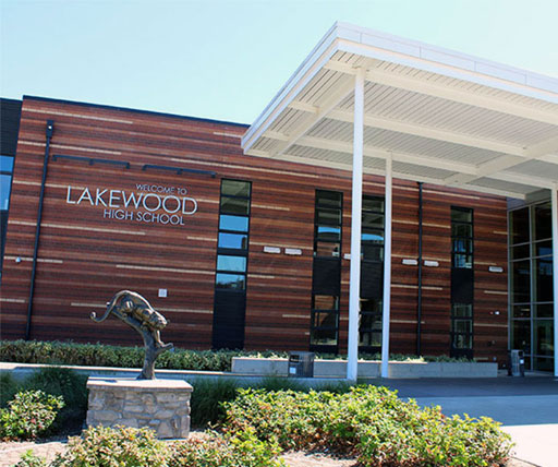
Anatomage Table Improves Learning Outcomes for Lakewood High School Students
At Lakewood High School in Long Beach, CA, science teacher Aaron Volkoff is using the Anatomage Table to make his Healthcare Analysis course more engaging. Students participate in interactive cadaver lab activities, utilizing the Table's case library and digital cadavers to understand real and pathological anatomy. This hands-on approach has significantly increased student interest and engagement. Read our case study to see how the Anatomage Table is transforming education at Lakewood High.