Anatomage Table – Google Ads [2025]
Shaping Tomorrow's Medical Education
We’d love to hear from you! Whether you have questions about our products, pricing, want to schedule a demo, or have other inquiries, reach out using the form and we will be in touch via email soon.
The Anatomage Table is the most advanced real-human-based medical education system. This state-of-the-art platform offers digitized human cadavers and superior medical learning tools, transforming medical education and training. By incorporating the Anatomage Table, institutions can enhance learning outcomes, lower laboratory costs, and establish their technological leadership.
Real Cadaveric Content
Offers digitized cadavers, real patient cases and simulation of real-tissue physiology.
Versatile Applications
Provides capabilities tailored to both education and training purposes.
Trusted By
Taking Education To The Next Level
Improve Outcomes
Research shows that using the Anatomage Table boosts learning outcomes. For instance, a study conducted with the University of Heidelberg found that this advanced tool improves test scores by an impressive 27%.
Save Cost & Time
The Anatomage Table reduces costs by replacing physical cadavers with digitized real human bodies, lowering lab maintenance fees and saving time in lab management.
Be a Tech Leader
By adopting the Anatomage Table, institutions can upgrade their labs with advanced technology, establishing themselves as leaders in medical education.
Main features
Anatomage Table features various life-size real human bodies in digital formats that provide an accurate representation and simulation of real 3D anatomy, physiology, and digital pathology.
Anatomage Bodies
Real Frozen Cadaveric Slices.
Anatomage Bodies are built from frozen cadaver slices, segmented from real human bodies donated for research. These slices underwent an intensive reconstruction process to re-create the cadaver’s pre-mortem form in a 3D digital format.
Intricate Vascular Systems.
The entire system of arteries, veins, and capillaries are fully traced and functionally connected, offering detailed insight with 3D anatomy.
Various Cadavers.
The Anatomage Body portfolio includes male, female, geriatric and pregnant cadavers.
High-Visualization.
3D anatomy details can be visualized up to 0.5 mm detail.
Annotated Structures.
Both female and male cadavers feature thousands of annotated structures. Students can select or locate specific anatomical structures from a comprehensive list.
Real Anatomy. Real Physiology.
Anatomage Bodies also demonstrate physiological functions similar to living bodies. Built from real cadaveric slices, they display the physical behaviors of real tissues during physiological processes, enabling students to accurately understand and visualize these functions.
Real-Tissue Simulation.
Our rendering technology integrates accurate physical motions with real tissues during functional processes, providing realistic physiological simulation.
Comprehensive Physiology.
Our physiological simulation tools include: Cardiac Motions, Kinesiology, Ocular Motions, General Pathways, Dental Arch, and Pregnancy & Delivery.
Complete Learning Solutions
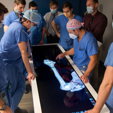
In-depth Clinical Database
- 1,600+ real-patient cases
- 1,100+ digital histology scans
- 77 photorealistic prosected scans
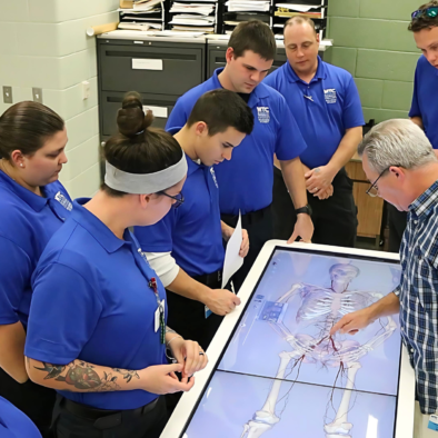
Clinical Procedure Simulation
- Specialized procedures include: Volumetric Dissection, Ultrasound, Craniotomy, Point to Point, Endoscopic View, and Catheterization
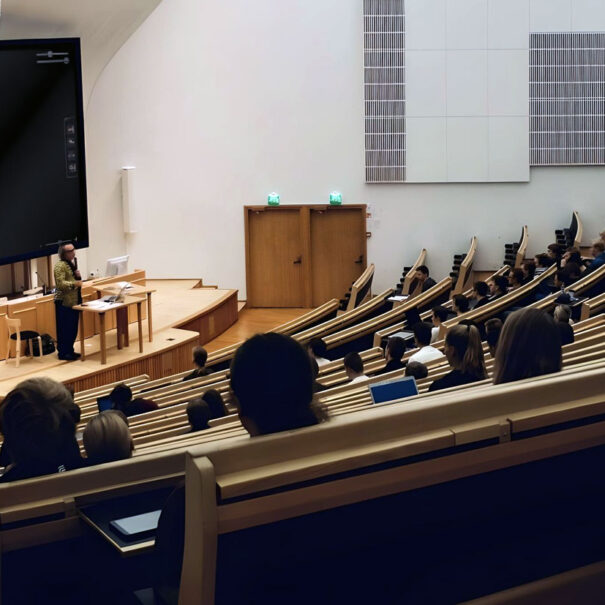
Learning Tools
- Customized quizzes
- Lecture tools
- 1,031 case-specific presets of anatomical structures
Clinical Applications
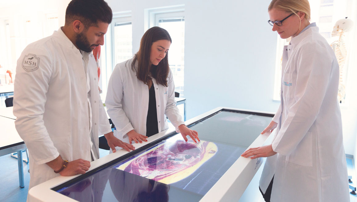
Diagnosis
Our Table lets you interpret, analyze, and examine patient scans with precision, utilizing state-of-the-art 3D DICOM rendering capabilities.
Surgical Training
Reduce surgical errors through virtual operative planning on Anatomage Bodies. Medical trainees and professionals can master surgical techniques and improve procedural accuracy in a risk-free setting.
Patient Communications
No more medical jargon. Patient pathologies are clearly displayed on an 84-inch touchscreen platform, enabling doctors to explain medical conditions more clearly.

University of Heidelberg Medical School
Anatomage Table Increased Student Scores by Up to 27%

In this case study, discover the results of an assessment conducted by researchers from the University of Heidelberg, the German Cancer Research Center, and the Karlsruhe Institute of Technology. They collaborated to evaluate the benefits of Anatomage Tables and cadaver CT scans on student learning of radiologic anatomy.
