Case Studies

University of Heidelberg Medical School
Discover how the Anatomage Table increased student scores in gross anatomy courses by up to 27%.

Tarrant County College
See why the Anatomage Table offers great benefits for anatomy courses in a community college setting.

Stanford University
Find out why the Anatomage Table received excellent reviews from Stanford program students.

The Harker School
Discover how this high school transformed their anatomy education with the Anatomage Table.

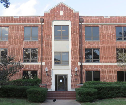
Prairie View A&M
Explore how the Anatomage Table helped promote spatial & conceptual learning of anatomy in biology courses.

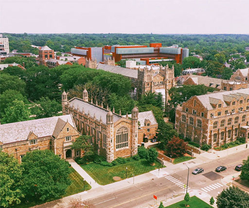
University of Michigan
See how this undergraduate nursing program successfully integrated the Anatomage Table into its curriculum.

University of Nebraska
Find out how the Anatomage Table helped increase exam scores and GPA for an imaging science program.

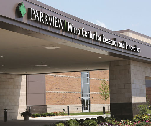
Parkview Mirro Advanced Medical Simulation Lab
Explore the Anatomage Table's impact in a simulation center setting.

Winthrop University
See how this university uses Anatomage technology to assess student learning outcomes in Anatomy & Physiology.

Samford University
Discover how the Anatomage Table helps physical therapy students excel at anatomy concepts.

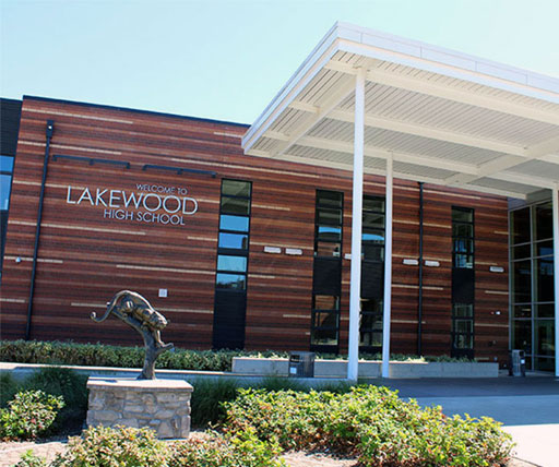
Lakewood High School
Find out how the Anatomage Table is increasing student engagement and improving learning outcomes.
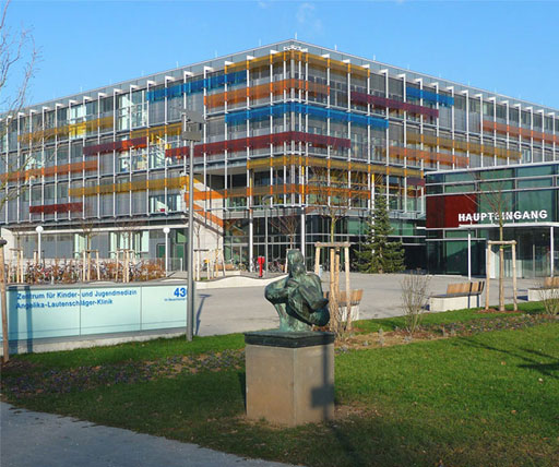
University of Heidelberg Medical School
Discover how the Anatomage Table increased student scores in gross anatomy courses by up to 27%.
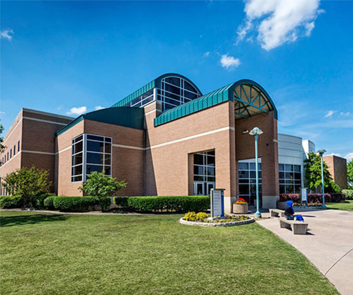
Tarrant County College
See why the Anatomage Table offers great benefits for anatomy courses in a community college setting.
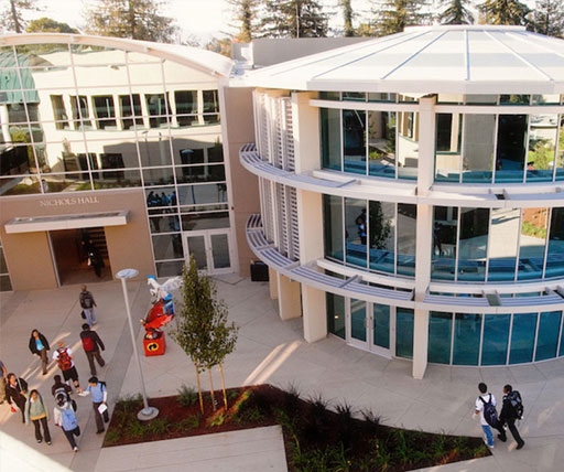
Stanford University
Find out why the Anatomage Table received excellent reviews from Stanford program students.
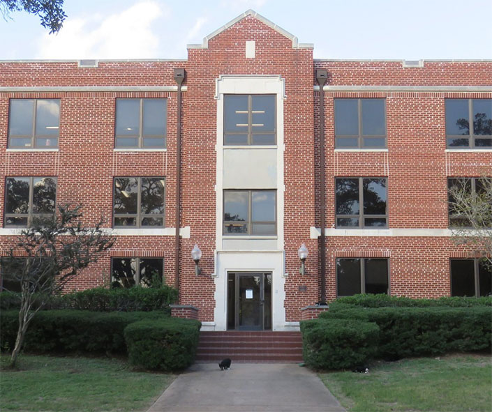
The Harker School
Discover how this high school transformed their anatomy education with the Anatomage Table.
Prairie View A&M
Explore how the Anatomage Table helped promote spatial & conceptual learning of anatomy in biology courses.
University of Michigan
See how this undergraduate nursing program successfully integrated the Anatomage Table into its curriculum.